Full Length Research Article - (2023) Volume 18, Issue 4
Relationship Between Sagittal Spinal Curvature And Incorrect Knee Position In School Age Children
Maha Ahmed Elsayed Soliman1*, Amira Mohamed Eltohamy2 and Mohamed Ali Elshafey2*Correspondence: Maha Ahmed Elsayed Soliman, Ph.D. Candidate, Al Ahrar Teaching hospital in Zagazig, Sharqia, Egypt, Email:
Abstract
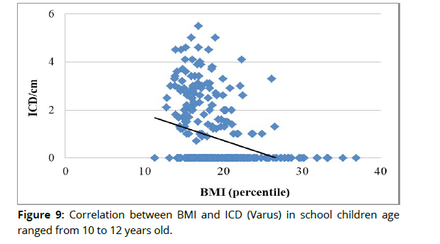
Objectives: To identify the relationship between sagittal spinal curvature and incorrect frontal knee position in school age children in Alsharqia, Egypt. Methods: 370 children of both genders were randomly selected from 10 Egyptian governmental schools in Alsharqia governorate aged from 10 to12 years old. The Saunders digital inclinometer was used to measure the patient's thoracic kyphosis and lumbar lordosis. Varus and valgus knees were diagnosed using sliding caliper in an upright position based on intercondylar distance with the feet placed together and intermalleolar distance with the knees placed together. Results: there was no significant relation between BMI and thoracic kyphosis, thoracic kyphosis and knee varus and valgus, lumber lordosis with knee varus and valgus (P>0.05). There is no significant relations between gender, BMI, thoracic kyphosis, lumber lordosis and valgus knee position (P>0.05). However, there was significantly negative correlation between gender and knee varus in school children (P<0.05). The direction of the relation between gender and knee varus increased in boys than girls. There were significantly positive correlations between BMI with lumber lordosis and knee valgus (IMD) (P<0.05) and negative correlation with knee varus (ICD) (P<0.05) in school children. Conclusion: Sagittal spinal curvature does not correlate with frontal knee position while BMI significantly correlates with lumber lordosis, knee valgus and knee varus position.
Keywords
spinal curvature. Incorrect knee position. School age children. Digital inclinometer. Sliding caliper
Introduction
Human health greatly benefits from maintaining an upright posture. Factors that have an impact on proper body posture fall into the categories of morphological, psychological, as well as environmental ones. Everyday habits, lifestyle, in addition to type of physical exercise engaged in, which may include different sports, all play a role1.
One of the most widespread but sometimes ignored health issues among children and teenagers is poor body posture. Uncorrected poor posture can lead to a variety of health problems, including impaired cardiorespiratory function, reduced lung vital capacity, degenerative bone & low back pain, and even internal organ displacement if left untreated2. One of the most widespread health issues across all age groups is poor posture and the musculoskeletal dysfunctions it induces. They impact around half of all children and adolescents3. Postural abnormalities have seen a 10–70% increase in prevalence over the previous decades due to changes in lifestyle. Lack of physical activity, Participating in Electronic Gaming, heavy backpacks, as well as persistent poor eating habits have been demonstrated in this regard4. Having a genu varum increases a person's chance of developing patellofemoral pain syndrome as well as osteoarthritis later in life because the force line shifts medially far from the joint centre, causing the medial joint reaction force to be nearly three and a half times that of the lateral compartment4. Both valgus as well as varus knees substantially overloading the knee joint, alter the patella position in the patellofemoral joint as well as increasing the risk of ligament damage5. The myofascial network suggests that there may be a connection between valgus as well as varus knee positions and the sagittal plane position of the spine, although this has not been well studied6. Postural defect prevention is a multifaceted approach to ensuring one's physical and mental well-being. Because of the rapid changes that occur in the body throughout puberty, poor posture is particularly prevalent in teenagers and children. This is because the habits formed at this time tend to last a lifetime7. As there is lack of available data regarding postural defects in primary school children in Egypt, there is an arising need to address this gap of knowledge. Therefore, the purpose of the current study was screening for postural defects in primary school children in Alsharqia Governorate, Egypt and determining the relationship between sagittal spinal curvature and incorrect frontal knee position in school age children. This would help understanding the mechanism of faulty posture and furthermore prevention of permanent postural defects.
Methods
Study design
From October 2022 until June 2023, a cross-sectional analysis was conducted. The Faculty of Physical Therapy at Cairo University, Egypt's Ethical Research Committee accepted the study (No:P.T.REC/012/003477). Each child's parent provided written informed permission after being briefed on the study's protocols.
Sample design
The sample design was a five stages cluster sample; 1st stage: select Al sharqia governorate from Egypt governorates, 2nd stage: select the largest 10 educational administrations from educational administrations. 3rd stage: select 10 primary schools from the largest10 educational administrations, one school from each educational administration. 4th stage: select children from grade five and grade six. 5th stage: random sampling of children from the different schools by choosing random identification number from the children list in each school and every odd number choose one child.
Subject
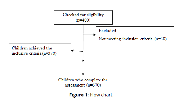
Four hundred children of both genders from 10 Egyptian governmental schools in Alsharqia governorate 40 child from each school participated in this study. As indicated in the flowchart in Fig. (1), 370 child were able to complete the evaluation after initially excluding 30 for not fulfilling the inclusion criteria.
The children were all between the ages of 10 and 12, all children were free from any medical disease, all children were studying at the Egyptian governmental schools in Alsharqia governorate and typically developing children from both sexes. Children were excluded if they have history of previous spine and lower extremities surgery, congenital deformity in lower extremities or trunk, neuromuscular disorders of the spine and lower extremities, trauma to the musculoskeletal system (such as a sprain or fracture) as well as children who participate regularly in sports (Figure 1).
Materials
1. Evaluation sheet for each child
2. Standardized weight and height measuring scale:
VI KI weighting scale ZT-220, Made in china, Serial Number: 498-930, Weights: 220 kilos.
3. Body mass index growth charts for boys and girls:
The BMI value was examined concerning the gender and age differences. The BMI status was categorised into four categories based on the attained percentile ranking obesity (BMI ≥ 95 percentile), overweight (BMI ≥ 85 percentile as well as <95 percentile), normal body mass (BMI <85 percentile as well as ≥ 5 percentile), in addition to underweight (BMI <5 percentile).
4. Marker: It was used to mark the first thoracic (T1) vertebra's spinous process, twelfth thoracic vertebrae (T12), fifth lumbar vertebra (L5), medial malleolus and medial femoral condyles on the children’s skin.
5. Digital inclinometer:
The degrees of kyphosis in the thoracic spine as well as lordosis in the lumbar spine were measured using a Saunders digital inclinometer (Baseline Digital Inclinometer, The Saunders Group Inc, Chaska, MN, USA).
6. Sliding caliper:
It was used to measure the degrees of knee valgus (intermalleolar distance) and varus (intercondylar distance). Caliper,9" SKU 78-1860, stainless steel handed, made in Germany had been used.
Procedures
All permissions which needed to assess the children at schools had been taken from The Directorate of education in Al Sharqia Governorate and the 10 educational administrations in Al Sharqia Governorate. Each child was assessed individually in a Calm and private room.
Weight, height and BMI
Weight and height were recorded for all children using weight and height measuring scale. The formula for determining BMI was as follows: weight (kg) / [height (m)]2.
Measurements of sagittal spinal curvature (thoracic kyphosis and lumber lordosis)
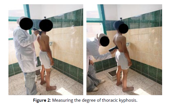
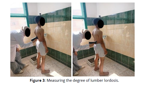
The measurements were taken while the subject was standing comfortably with their head held in upright position, their arms at their sides, extended knees, as well as feet shoulder-width apart8. The spinous process of the T1, T12, as well as L5 were highlighted on the children’s skin9. Saunders digital inclinometer (Baseline Digital Inclinometer, The Saunders Group Inc, Chaska, MN,USA) was used to measure kyphosis and lordosis in the thoracic and lumbar regions. The inclinometer was set to cradle the spinous processes and were gently but firmly pressed into the spaces between the spine10. Initially, the inclinometer was positioned on (T1) and calibrated to 0º. The inclinometer was subsequently put on the (T12), the grades of the thoracic curve have been recorded. When put on the (T12), the inclinometer was calibrated to 0 once more the inclinometer was then positioned at the third point (L5) as well as the grades for the lumber curve have been recorded10. The thoracic kyphosis and lumbar lordosis measures displayed in Fig (2,3) (Figures 2 & 3).
The normal standing thoracic curve reference is decreased kyphosis or hypokyphosis lower than 20 degrees, normal kyphosis is between 20 and 40 degrees, while hyperkyphosis is greater than 40 degrees. Regarding lumbar curvature, the following classifications were utilized: hypolordosis lower than 20 degrees, normal lumbar is between 20 and 40 degrees, hyperlordosis greater than 40 degrees11.
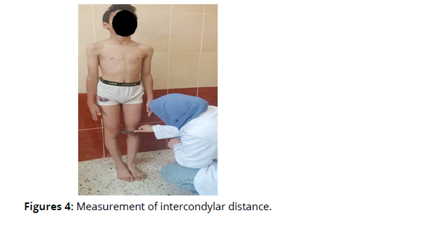
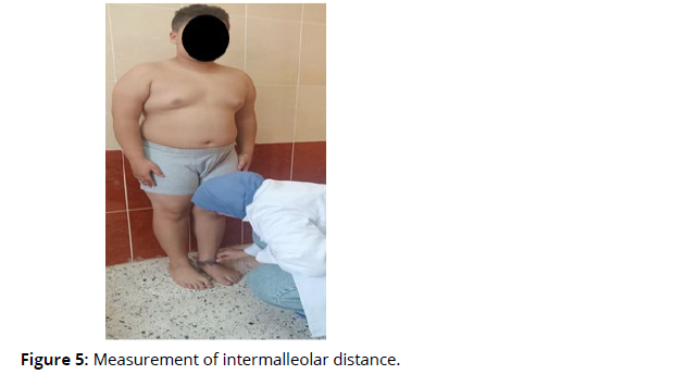
Measurements of frontal knee position (knee varus and valgus) The children were examined while standing barefoot with their knees completely extended. Medial malleolus and medial femoral condyles had been marked on the children’s skin. The intercondylar distance (ICD) was measured with the feet together, medial malleoli of the ankles contacting to identify a valgus knee. The intermalleolar distance (IMD) was measured with the knees together, with the two medial femoral condyles contacting each other, to identify valgus knees5. A sliding caliper (Caliper,9" SKU 78-1860) was used for the measurements. The measurements of ICD as well as IMD represented in Fig (4,5) (Figures 4&5).
Both valgus and varus knees are considered abnormal in children of the age range used in the study. Therefore, readings of IMD and ICD greater than 2.5 cm will be regarded as abnormal. The caliper was chosen because it was easy to use, accurate, and there was no need to subject the children in the investigation to X-rays5.
Statistical analysis
The Shapiro-Wilk test was employed to determine whether or not the study variables followed a normal distribution. This study's statistical analysis was performed in SPSS for Windows, version 25. (SPSS, Inc., Chicago, IL).
Quantitative descriptive statistics data comprising the mean as well as standard deviation and qualitative descriptive statistics data including frequency had been used. Bi-variate correlation coefficient was conducted to compute the relation and direction among the study variables. The results of every statistical test were statistically significant at the 0.05 level (P ≤ 0.0).
Results
1. Homogeneity test
The Shapiro-Wilk test was employed to determine whether or not the study variables followed a normal distribution. This test revealed that data of age (P=0.253), weight (P=0.249), height (P=0.269), BMI (P=0.273), thoracic kyphosis/ degree (P=0.207), lumber lordosis/degree (P=0.191), ICD/cm (P=0.361), and IMD/cm (P=0.378) were normally distributed (P>0.05).
2. Descriptive statistic for children of all measured variables:
A total of 370 children of both sexes took part in the study with average age, weight, and height were 10.93±0.57, 39.83±10.33, and 185.02 ±60.96, respectively (Table 1). Descriptive statistic for children of all measured variables presented in (Table 1).
| Variables | Category | All administrations |
|---|---|---|
| Number | N | 370 |
| Age (years) | Mean ± SD | 10.93 ±0.57 |
| Weight (kg) | Mean ± SD | 39.83 ±10.33 |
| Height (cm) | Mean ± SD | 185.02 ±60.96 |
| Grade | 5th grade | 215 (58.10%) |
| 6th grade | 155 (41.90%) | |
| Gender | Boys | 234 (63.20%) |
| Girls | 136 (36.80%) | |
| BMI | Underweight | 31 (8.30%) |
| Healthy weight | 237 (64.10%) | |
| Overweight | 64 (17.30%) | |
| Obesity | 38 (10.30%) | |
| Thoracic kyphosis / degree | Hypokyphosis <20◦ | 1 (0.30%) |
| Normal kyphosis 20◦ - 40◦ | 135 (36.50%) | |
| Hyperkyphosis >40◦ | 234 (63.20%) | |
| Lumber lordosis / degree | Hypolordosis<20◦ | 106 (28.60%) |
| Normal lumbar 20◦ - 40◦ | 249 (67.30%) | |
| Hyperlordosis >40◦ | 15 (4.10%) | |
| Frontal knee position (ICD,IMD) | Valgus ≥2.5 | 55 (14.90%) |
| Normal <2.5 | 251 (67.80%) | |
| Varus ≥2.5 | 64 (17.30%) |
Quantitative data (age, weight, and height) are expressed as mean ±standard deviation.
Qualitative data (grade, gender, BMI, thoracic kyphosis, lumber lordosis, ICD, and IMD) are expressed as frequency (percentage).
3. Correlation between gender with BMI, thoracic kyphosis, lumber lordosis, ICD, and IMD in school children
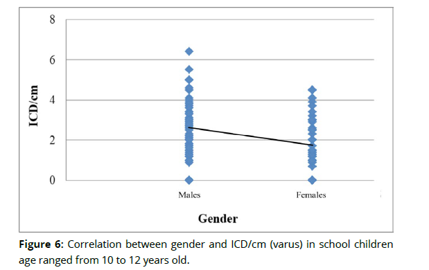
Bi-variate Spearman correlation coefficients were computed between gender with BMI, thoracic kyphosis, lumber lordosis, ICD, and IMD in school children (Table 2).Correlation between gender and ICD/cm (varus) in school children presented in Fig (6). (Table 2 & Figure 6)
| Items | Gender (n=370) | ||
|---|---|---|---|
| r - value | P-value | Significance | |
| BMI | 0.085 | 0.101 | NS |
| Thoracic kyphosis | -0.014 | 0.782 | NS |
| Lumber lordosis | 0.058 | 0.269 | NS |
| ICD (Varus) | -0.112 | 0.032* | S |
| IMD (Valgus) | 0.060 | 0.253 | NS |
| r: Spearman-rank correlation coefficient value; P-value: probability value; *Significant: (P<0.05); NS: non-significant. |
|||
4. Correlation between BMI with thoracic kyphosis, lumber lordosis, ICD, and IMD in school children
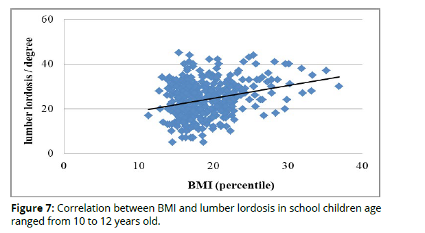
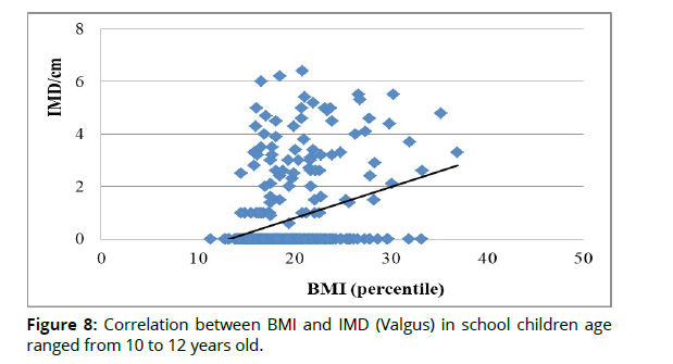
Bi-variate Spearman correlation coefficients were computed between BMI with thoracic kyphosis, lumber lordosis, ICD, and IMD in school children (Table 3).The direction of the relations between BMI with lumber lordosis, valgus(IMD) and varus(ICD) presented in Fig (7,8,9) (Table 3 & Figures 7,8,9).
| Items | Gender (n=370) | ||
|---|---|---|---|
| r - value | P-value | Significance | |
| BMI | 0.078 | 0.135 | NS |
| Thoracic kyphosis | 0.186 | 0.0001* | NS |
| Lumber lordosis | -0.253 | 0.0001* | NS |
| ICD (Varus) | 0.245 | 0.0001* | S |
| IMD (Valgus) | 0.060 | 0.253 | NS |
| r: Spearman-rank correlation coefficient value; P-value: probability value; *Significant: (P<0.05); NS: non-significant. |
|||
5. Correlation between thoracic kyphosis with ICD and IMD in school children
Bi-variate Spearman correlation coefficients were computed between thoracic kyphosis with ICD and IMD in school children (Table 4).
| Items | Gender (n=370) | ||
|---|---|---|---|
| r - value | P-value | Significance | |
| ICD (Varus) | 0.083 | 0.113 | NS |
| IMD (Valgus) | 0.004 | 0.935 | NS |
| r: Spearman-rank correlation coefficient value; P-value: probability value; *Significant: (P<0.05); NS: non-significant. |
|||
6. Correlation between lumber lordosis with ICD and IMD in school children
Bi-variate Spearman correlation coefficients were computed between lumber lordosis with ICD and IMD in school children (Table 5).
| Items | Gender (n=370) | ||
|---|---|---|---|
| r - value | P-value | Significance | |
| ICD (Varus) | 0.024 | 0.649 | NS |
| IMD (Valgus) | 0.013 | 0.804 | NS |
| r: Spearman-rank correlation coefficient value; P-value: probability value; *Significant: (P<0.05); NS: non-significant. |
|||
Discussion
This research was conducted to investigate if there is a relationship between the degree of thoracic kyphosis and genu valgus and varus, the degree of lumber lordosis, and genu valgus and varus, the body mass index and genu valgus and varus, the body mass index and thoracic kyphosis and lumber lordosis in school age children. Screening assessment for abnormalities of sagittal spinal curvature (kyphosis and lordosis) and frontal knee position (varus and valgus) was conducted. Descriptive statistics of the current study revealed that the percentage of children BMI categories underweight, healthy weight, overweight, and obesity were (8.30%), (64.10%), (17.30%), and (10.30%). The percentage of children frontal knee position categories valgus, normal, and varus were (14.90%), (67.80%), and (17.30%), respectively. These results are in agreement with those found in the of Shapouri12, who said 1,450 students were chosen at random from a pool of 20,000 school-aged children. In the population that was examined, 17.7 % had genu valgum, 24.2% had genu varum, and 13.38% had flat feet. Children are more likely to suffer from the common postural abnormality known as genu varum. In a study of Brazilian children ages 7-12, Ciaccia et al. 13 found that 7.1% of them had knee valgus. Souza et al. identified a substantially greater incidence of valgus knee (56%) in another Brazilian study; nevertheless, over 60% of the investigated group was overweight or obese, which may have significantly impacted the results14. In the current study the percentage children thoracic kyphosis categories hypokyphosis, normal kyphosis, hyperkyphosis were (0.30%), (36.50%), and (63.20%), respectively. The percentage of children lumber lordosis categories hypolordosis, normal lumbar, hyperlordosis were (28.60%), (67.30%) and (4.10%), respectively. Two-thirds of children aged 10 to 12 were found to have abnormal spinal alignment in the sagittal plane, as reported by Agnieszka et al.5. These children also tended to have frontal knee positions which are out of alignment. Approximately 40 % had incorrect frontal knee posture. When Katarzyna et al. 2 examined a group of 888 children between the ages of 7 and 12, they found that the prevalence of rounded thoracic kyphosis, rounded lumbar lordosis, and flat back was significantly lower than in our study (7.1%, 6.6% and 1.8%, respectively). Nonetheless, this research was conducted in a different way (visual assessment of body posture), that might account for the differences. A study by Protić et al. 15 including a total of 55 children aged 10-13 years old showed that lordosis affects 67.3% of children, kypho-lordosis affects 40.5%, as well as kyphosis affects 32.8% of children. Straight backs were found in 5.5%. The results of correlational analysis of the recent research revealed that no substantial relations among gender with BMI, thoracic kyphosis, lumbar lordosis, and IMD in school children(P>0.05). In contrast, a substantial negative correlation was found among gender as well as ICD among schoolaged children (P<0.05). The direction of the relation between gender and ICD (varus) increased in boys than girls, which comes in line with the findings of the study by Furian et al .16 who used the rasterstereography technique to study 345 children aged 10-11 and found no correlation between gender and thoracic kyphosis severity. Furthermore, Erkan et al. 17 argue that there is no difference in the severity of thoracic kyphosis based on gender, whereas Lau et al. 18 showed a 2.4% incidence of abnormal kyphosis among male students as well as a 1.2% prevalence among female students. In the present study, the results of correlational analyses revealed that no significant relation between BMI with thoracic kyphosis in school children(P>0.05). However, there were significantly positive correlations among BMI with lumber lordosis and IMD (valgus knee) and negative correlation with ICD (varus knee) in school children (P<0.05). These results are in accordance with those of Molina et al. 19 who found that the children who were overweight were more likely to have lumbar hyperlordosis, genu valgum, flatfoot, as well as other joint malalignments than their normal weight peers. When a person has a (BMI) that is too high, the spine is put under unnecessary stress. This causes the muscles as well as other structures of the spine to become functionally overloaded Shohat et al.20. Micha et al.21 found that the prevalence of limb deformities was highest in children who were overweight or obese, and lowest among children who were normal weight or underweight, supporting our own findings. Valgus knee was the most frequent postural abnormality observed in children who were overweight or obese. Agnieszka et al. 22 found that one's weight is a determining factor in how their trunks are positioned, and these findings are consistent with those of the present investigation. The lumbar lordosis was found to be impacted by both BMI and fat percentage. Other investigators, such as Go'rniak et al. 23, have also noticed a correlation between extreme body weight as well as the lumbar lordosis's shape. The current study agrees with previous studies on students in southern Africa that linked an increment in BMI to an increment in lumbar hyperlordosis 24. Similar to our findings, Katarzyna et al. 2 found that obesity is linked to poor posture in children and adolescents between the ages of 7 and 12, with valgus knees being the most frequent postural deviation among obese children and adolescents. However, there is limited researches studying the relationship between sagittal spinal curvature and frontal knee position. The results of correlational analyses in the present study revealed that no significant relations between lumber lordosis with ICD and IMD in school children which comes in accordance with the results of the study by Agnieszka et al.5 which revealed that the incidence of varus knees does not appear to be related to the shape of the lumbar spine, since the degree of lumbar lordosis did not change between groups with valgus, varus, or correctly positioned knees. Along with the present study, there are multiple concepts regarding myofascial chains, the most well-known of which are the hypotheses of Stecco25 and Myers26 in each of these, connections were observed between the tissues of the trunk and the muscles and fascia of the lower limbs. This implies that these structures interact, where it states that changes in the position of one section of the body caused by overloading the myofascial structures alter the load on the segments above as well as below, but none of these theories adequately illustrates the impact of lower limb positioning on the position of the spine. Also, The results of correlational analyses in the present study revealed that no substantial relations among thoracic kyphosis with ICD(varus knee) and IMD (knee valgus) in school children while the study by Agnieszka et al. 5 reported substantial correlation among flat thoracic kyphosis as well as valgus knee position in addition to between varus knees as well as increased thoracic kyphosis, Valgus knees are more frequent in patients having flat thoracic kyphosis, The value of the correlation coefficient indicates that the relationship, despite being significant based on the p-value, was weak.
Study limitations:
The research methods used certainly, X-rays would be the gold standard for evaluating the position of the spine as well as knees, but there was no necessity to expose children to the harmful effects of radiation.
Lack of cooperation of some school managers and children's caregivers and refusal to participate in the study (for girl’s children) due to traditional thoughts. So, it is recommended that postural evaluation and screening for postural defects in primary school children needs to be a part of their health routine and using the obtained results to construct a structured evaluation based rehabilitation program to prevent or correct possible spine and knee abnormalities.
Conclusion
The study concluded that Sagittal spinal curvature does not correlate with frontal knee position while BMI significantly correlate with lumber lordosis, knee valgus and knee varus position. So, further researches with larger sample size are needed in order to better clarify the relationship between sagittal spinal curvature and abnormal knee position
References
Walicka CV, Katarzyna , Szeliga E , Guzik A , Mrozkowiak MB , Niewczas M , Ostrowski PB, and Tabaczek BI. Evaluation of Anterior-Posterior Spine Curvatures and Incidence of Sagittal Defects in Children and Adolescents Practicing Traditional Karate . BioMed Research International 2019 :Volume, Article ID 9868473, 9 pages https://doi.org/10.1155/2019/9868473.
Katarzyna MP ,Barbara SW,Tomasz K ,Anna S ,Alicja K ,Jarosław W and Małgorzata KW. Prevalence of incorrect body posture in children and adolescents with overweight and obesity Eur J Pediatr 2017 ;176(5):563-572. doi: 10.1007/s00431-017-2873-4. Epub.
Ludwig O. Interrelationship between postural balance and body posture in children and adolescents. J Phys Ther Sci 2017; 29(7):1154–8. https://doi.org/10.1589/jpts.29.1154 PMID: 28744036.
Sahar SG , Sami EE , Abdelmoniem AY ,Asmaa MA and Nashwa I H. Prevalence of lower limb deformities among primary school students: Egyptian Rheumatology and Rehabilitation 2021 :48:34 https://doi.org/10.1186/s43166-021-00082-1.
Agnieszka JS , Michał F , Eliza S , Renata B , Katarzyna W. Relationship between frontal knee position and the degree of thoracic kyphosis and lumbar lordosis among 10-12-year-old children with normal body weight. PLoS ONE 2020 :15(7): e0236150.
Shah S and Bhalara A. Myofascial Release. Int J Heal Sci Res 2012:[Internet].
Dorota L , Wojciech C , Marta N.The problem of postural defects in children and adolescents and the role of school teachers and counselors in their prevention. Scientific Review of Physical Culture 2014 : volume 4, issue 4.
Alena C ,Erika Z ,Ľubomír Š, Marián U, José M M . Spinal curvature in female and male university students with prolonged bouts of sedentary behavior .Research Square 2022 : DOI: https://doi.org/10.21203/rs.3.rs-1989231/v1.
Angelica GD , Maria TMR, Antonio C ,Alba AS, Pillar S . Sagittal Spinal Morphotype Assessment in Dressage and Show Jumping Riders. J. Sport Rehabil 2019 : 18;29(5):533-540. doi: 10.1123/jsr.2018-0247.
López MPA, Alacid F and Rodríguez GPL. Comparison of sagittal spinal curvatures and hamstring muscle extensibility among young elite paddlers and non-athletes. Int. Sport Med. J. 2010: 11, 301–312.
Fernando SM , Mónica CD, María TMR, Olga RF, Alba AS, Antonio C, Pilar A and Pilar S B (2020). Classification System of the Sagittal Integral Morphotype in Children from the ISQUIOS Programme (Spain) Int. J. Environ. Res. Public Health, 17, 2467; doi:10.3390/ijerph17072467 www.mdpi.com/journal/ijerp.
Shapouri J, Mohammad A , Marzieh A , Abolfazl I , Robabeh A and Silva H Prevalence of Lower Extremities’ Postural Deformities in Overweight and Normal Weight School Children Iran J Pediatr. 2019; 29(5):e89138.
Ciaccia MCC, Pinto CN, Golfieri F d C, Machado TF, Lozano LL, Silva JMS, et al. Prevalência de genuvalgo em escolas públicas do ensino fundamental na cidade de santos (sp), brasil. Rev Paul Pediatr. 2017;35:443–7.
Souza AA, Ferrari GL De M, Silva Júnior JP Da, Silva LJ Da, Oliveira LC De, Matsudo VKR. Association between knee alignment, body mass index and physical fitness variables among students: a cross-sectional study. Rev Bras Ortop (English Ed. 2013;48:46–51.
Protić Gava, B., Šćepanović, T., Jevtić, N., and amp; Kadović, V. Frequency of postural disorders in the sagittal plane of younger school age. Activities in Physical Education & Sport 2011 . 2 (6), 151-156.
Furian TC, Rapp W, Eckert S, Wild M, Betsch M. Spinal posture and pelvic position in three hundred forty-five elementary school children: a rasterstereographic pilot study. Orthop Rev (Pavia) [Internet]. 2013; 5(1):29–33
Erkan S, Yercan HS, Okcu G, O¨ zaip RT. The influence of sagittal cervical profile, gender and ageon the thoracic kyphosis. Acta Orthop Belg. 2010; 76:675–80. PMID: 21138225.
Lau KT, Cheung KY, Chan KB, Chan MH, Lo KYand Chiu TT. Relationships between sagittal postures of thoracic and cervical spine, presence of neck pain, neck pain severity and disability. Man Ther 2010;15:457-62.
Molina-Garcia, P.; Miranda-Aparicio, D.; Ubago-Guisado, E.; Alvarez-Bueno, C.; Vanrenterghem, J.; Ortega, F.B. The Impact of
Childhood Obesity on Joint Alignment: A Systematic Review and Meta-Analysis. Phys. Ther. 2021, 101, pzab066. .
Shohat, N.; Machluf, Y.; Farkash, R.; Finestone, A.S.; Chaiter, Y. Clinical Knee Alignment among Adolescents and Association
with Body Mass Index: A Large Prevalence Study. Isr. Med. Assoc. J. IMAJ 2018, 20, 75.
Michał Brzeziński1 , Zbigniew Czubek , Aleksandra Niedzielska , Marek Jankowski , Tomasz Kobus and Zbigniew Ossowski. Relationship between lower-extremity defects and body mass among polish children: a cross-sectional study Brzeziński et al. BMC Musculoskeletal Disorders ,2019.20:84 https://doi.org/10.1186/s12891-019-2460-0.
Agnieszka Jankowicz-Szyman´ska, PhD, Marta Bibro, MA, Katarzyna Wodka, MA, and Eliza Smola, MA. Does Excessive Body Weight Change the Shape of the Spine in Children? CHILDHOOD OBESITY, July 2019, Volume 15, Number 5.
Go´rniak K, Lichota M, Pop1awska H, et al. Postawa cia1a ch1opco´w wiejskich z niedoborem i nadmiarem tkanki t1uszczowej w organizmie [in polish] [Body posture of rural boys with deficiency or excess of body fat]. Rocznik Lubuski 2014;40:163–176.
Malepe MM, Goon DT, Anyanwu FC, et al. The relationship between postural deviations and body mass index among university students. Biomed Res 2015;26:437–442.
Stecco C. Functional Atlas of the Human Fascial System. Elsevier Health Sciences.; 2014.
Myers TW. Anatomy Trains: Myofasziale Leitbahnen (fu¨r Manual-und Bewegungstherapeuten). Elsevier, Urban & Fischer Verlag; 2015.
